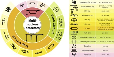
It has always been of considerable interest to study the nuclear magnetic resonance response of multiple nuclei simultaneously, whether these signals arise from internuclear couplings within the same molecule, or from uncoupled nuclei within sample mixtures. The literature contains numerous uncorrelated reports on techniques employed to achieve multi-nuclear NMR detection. This paper consolidates the subset of techniques in which single coil detectors are utilized, and highlights the strengths and weaknesses of each approach, at the same time pointing the way towards future developments in the field of multi-nuclear NMR. We compare the different multi-nuclear NMR techniques in terms of performance, and present a guide to NMR probe designers towards application-based optimum design. We also review the applicability of micro-coils in the context of multi-nuclear methods. Micro-coils benefit from compact geometries and exhibit lower impedance, which provide new opportunities and challenges for the NMR probe designer.
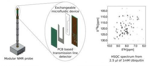
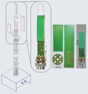
Microfluidic NMR spectroscopy can probe chemical and bio-chemical processes non-invasively in a tightly controlled environment. We present a dual-channel modular probe assembly for high efficiency microfluidic NMR spectroscopy and imaging. It is compatible with a wide range of microfluidic devices, without constraining the fluidic design. It collects NMR signals from a designated sample volume on the device with high sensitivity and resolution. Modular design allows adapting the detector geometry to different experimental conditions with minimal cost, by using the same probe base. The complete probe can be built from easily available parts. The probe body mainly consists of prefabricated aluminium profiles, while the probe circuit and detector are made from printed circuit boards. We demonstrate a double resonance HX probe with a limit of detection of 1.4 nmol s−1/2 for protons at 600 MHz, resolution of 3.35 Hz, and excellent B1homogeneity. We have successfully acquired 1H-13C and 1H-15N heteronuclear correlation spectra (HSQC), including a 1H-15N HSQC spectrum of 1 mM 15N labeled ubiquitin in 2.5 μl of sample volume.
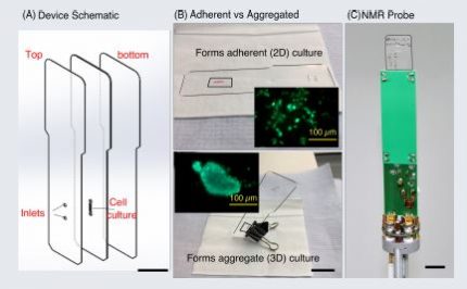
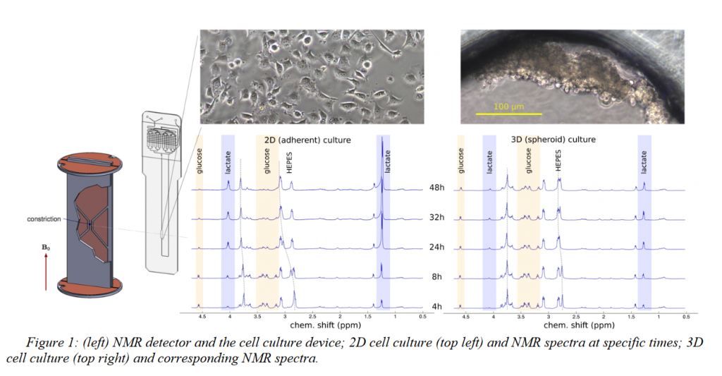
Time-lapse NMR spectroscopy was achieved up to 48 hours with MCF-7 cells, cultured in a microfluidic device of volume 2.5 l
Cells were cultured as an attached monolayer (2D) or an aggregated spheroid (3D) using same device design
Cell viability analysis was performed at the end of the NMR experiments
Manvendra Sharma, Bishnubrata Patra, William Hale and Marcel Utz
University of Southampton
ENC 2019
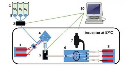
We present a novel microfluidic-NMR platform to combine high sensitivity NMR with microfluidics to perform generic NMR experiments on live systems under tightly controlled environment through microfluidics.
The current setup has already been used for a number of applications including microimaging of mouse liver tissue slices, heteronuclear 2D-NMR correlation experiments on proteins, parahydrogen induced hyperpolarisation, droplet NMR, reaction monitoring, and metabolic studies of mammalian cells.
Bishnubrata Patra, Manvendra Sharma, William Hale, Marcel Utz
University of Southampton
MicroTAS 2018
ABSTRACT
NMR metabolomics, the study of cellular metabolism, provides a new path to understand cellular response in vitro. In this article, we performed a real-time monitoring of adherent vs. spheroid cell culture using NMR metabolomics. The difference in lactate production provides a clear indication of different metabolic rates in spheroid compared to adherent cell culture. The developed microfluidic–NMR technique could be applicable towards real-time monitoring of a variety of ex-vivo biological samples, including tissue or organ on a chip.
Three-dimensional cell culture models (spheroids) provide valuable additional insight into the behavior of cancer cells since it mimics the features such as cell-cell contact and hindrance of metabolic diffusion which are not present in traditional cell culture.1On the other hand, NMR spectroscopy can provide in-situ monitoring of metabolomics even at microliter volume.2In this work, we used a novel integrated transmission-line NMR probe to directly compare the metabolomic activity of MCF-7 cells in adherent (2D) and spheroid (3D) culture. Microfluidic cell culture device of volume 2.5 μl volume was coated with fibronectin and with pluronic respectively to produce 2D and 3D cell cultures. Metabolomics observation was done up to 48 hours using NMR spectroscopy, followed by viability analysis. Clear differences in metabolomic activity were found, the spheroid (3D) showed much lower consumption of glucose, production of lactate, and acidosis compared to the attached (2D) culture.
EXPERIMENTAL
Microfluidic devices were made from thermally bonded PMMA layers. The cell culture chamber was coated overnight with fibronectin (1 mg/ ml) orpluronic (10 mg/ml). A suspension of 2.5 μl MCF-7 Cells (1000 cells/μl) in DMEM culture medium was introduced into each device. The devices were kept inside a cell culture incubator at 37° C and with 5% CO2. 1H NMR spectra were recorded using a 600 MHz spectrometerforboth2D and 3D cell culture devices and controls (without cells) periodically at 4, 8, 24, 32, and 48 hours. Cell viability was analyzed on the same devices using Live/Dead cell imaging kit [life technologies]and fluorescence microscopy.
RESULTS AND DISCUSSION
In this work, we used the equal number of cells with the equal volume of culture medium and the same culture environment to compare the metabolic activity of 2D vs.3D cultured MCF-7 cells by NMR spectroscopy. Our results showed that aggregated cells are metabolically less active and more viable after 48 hours compared to the attached cells
CONCLUSION
Figure 1 shows the 1HNMR spectra of 2D and 3D cultures up to 48 hours. 2D cells already started to produce more lactate compared to the 3D cell culture after 4 hours. At 48 hours 2D cell culture produces enough lactate to significantly shift the HEPES buffer resonances and consumed almost all the glucose. By contrast, 3D cells produced significantly less lactate. NMR spectra obtained in this way are of significant quality for unsupervised peak identification and metabolites present could be identified by mining the human metabolome database(figure 2). Figure 3 shows the viability analysis of 2D and Spheroid after 48 hours. The live stain (Green) indicates the healthy cells, whereas dead cells are stained as red. After 48 hours, 2D medium got enough acidified that cells are detaching, whereas cells survived strongly as spheroid.
ACKNOWLEDGEMENTS
This research project is funded by the horizon 2020 Framework program of the European Union.
REFERENCES
[1] F. Pampaloni, E. G. Reynaud, E.H.K. Stelzer, “The third dimension bridges the gap between cell culture and live tissue”. Nat Rev Mol Cell Biol. 8, 839, 2007
[2] G. Finch, A. Yilmaz, M. Utz, “An optimized detector for in-situ high-resolution NMR in microfluidic devices”. J. Mag. Res.262, 73-80, 2016
[3] D.S. Wishart, Y.D. Feunang, A. Marcu, A.C. Guo K. Liang, “ HMDB 4.0-The human metabolome database for 2018”. Nucleic Acids.Res.Jan 4; 4 6(D1): D608-17, 2018
Mazin Jouda, Erwin Fuhrer, Pedro Silva, Jan G. Korvink, and Neil MacKinnon
Karlsruhe Institute of Technology
ENC 2019
ABSTRACT
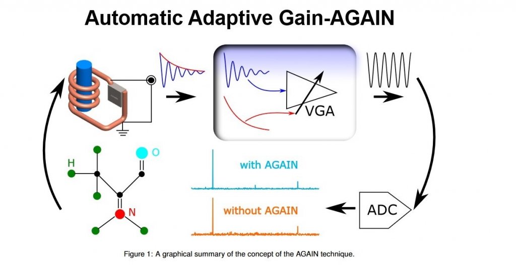
For an efficient NMR acquisition, an NMR spectrometer usually offers the option of automatically adjusting the receiver gain. The adjustment in this case is driven by the maximum amplitude of the FID, and once the receiver gain is set to a certain value, then it cannot be changed during the acquisition. But since the NMR signal is decaying by nature, only the first few points of the FID will be acquired with the full resolution of the analog-to-digital converter (ADC), whereas the rest of the FID will be acquired with a constantly decreasing resolution.
In this contribution we describe an innovative closed-loop control mechanism to automatically adapt the receiver gain over the acquisition time such that all of the FID points are digitized with the full ADC resolution [1]. A high performance digital lock-in amplifier was employed to extract the envelope of the FID, process it, and feed it back to control a variable-gain amplifier (VGA) inserted in the signal chain. As the FID decays the VGA gain and thus the overall receiver gain increases,even if the FID increases again due to beating for example, the VGA gain is adapted ensuring that no ADC overflow occurs.Additionally, the reported method includes a mechanism to precisely monitor the gain change over time. This is carried out via a pilot signal that propagates together with the NMR signal through the VGA. Knowledge of the gain modulation is of course essential to recover the NMR signal after the ADC stage.
The method, which we call AGAIN (Automatic Adaptive Gain), was verified experimentally. The results showed an SNR enhancement factor of 2.64 when AGAIN was applied to a custom spectrometer, while a maximum SNR enhancement factor of 1.45 was observed when applied to a commercial spectrometer. The AGAIN technique requires minimal additional hardware, which makes it general and thus can be readily combined with any commercial spectrometer.
REFERENCES
[1] M. Jouda, E. Fuhrer, P. Silva, J. G. Korvink, and N. MacKinnon. “Automatic adaptive gain for magnetic resonancesensitivity enhancement”. In:Analytical Chemistry (2019).
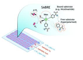
Complex mixtures, commonly encountered in metabolomics and food analytics, are now routinely measured by nuclear magnetic resonance (NMR) spectroscopy. Since many samples must be measured, onedimensional proton (1D 1H) spectroscopy is the experiment of choice. A common challenge in complex mixture 1H NMR spectroscopy is spectral crowding, which limits the assignment of molecular components to those molecules in relatively high abundance. This limitation is exacerbated when the sample quantity itself is limited and concentrations are reduced even further during sample preparation for routine measurement. To address these challenges, we report a novel microfluidic NMR platform integrating signal enhancement via parahydrogen induced hyperpolarisation. The platform simultaneously addresses the challenges of handling small sample quantities through microfluidics, the associated decrease in signal given the reduced sample quantity by Signal Amplification by Reversible Exchange (SABRE), and overcoming spectral crowding by taking advantage of the chemosensing aspect of the SABRE effect. SABRE at the microscale is enabled by an integrated PDMS membrane alveolus, which provides bubble-free hydrogen gas contact with the sample solution. With this platform, we demonstrate high field NMR chemosensing of microliter sample volumes, nanoliter detection volumes, and micromolar concentrations corresponding to picomole molecular sensitivity.
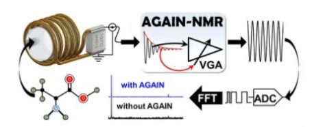
The decaying nature of magnetic resonance (MR) signals results in a decreasing signal-to-quantization noise ratio (SQNR) over the acquisition time. Here we describe a method to enhance the SQNR, and thus the overall signal-to-noise ratio (SNR), by dynamically adapting the gain of the receiver before analog-to-digital conversion (ADC). This is in contrast to a standard experiment in which the gain is fixed for a single data acquisition and is thus adjusted only for the first points of the signal. The gain adjustment in our method is done automatically in a closed loop fashion by using the envelope of the MR signal as the control signal. Moreover, the method incorporates a robust mechanism that runs along with signal acquisition to monitor the gain modulation, enabling precise recovery of the signals. The automatic adaptive gain (AGAIN) method requires minimal additional hardware and is thus general and can be implemented in the signal path of any commercial spectrometer system. We demonstrate an SNR enhancement factor of 2.64 when applied to a custom spectrometer, while a factor of 1.4 was observed when applied to a commercial spectrometer.