Glassy carbon microelectrodes minimize induced voltages, mechanical vibrations, and artifacts in magnetic resonance imaging

Surabhi Nimalkar, Erwin Fuhrer, Pedro Silva, Tri Nguyen, Martin Sereno, Sam Kassegne and Jan Korvink

Microsystems and Nanoengineering, 2019

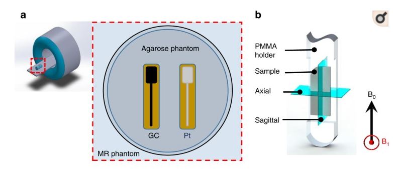
The recent introduction of glassy carbon (GC) microstructures supported on flexible polymeric substrates has motivated the adoption of GC in a variety of implantable and wearable devices. Neural probes such as electrocorticography and penetrating shanks with GC microelectrode arrays used for neural signal recording and electrical stimulation are among the first beneficiaries of this technology. With the expected proliferation of these neural probes and potential clinical adoption, the magnetic resonance imaging (MRI) compatibility of GC microstructures needs to be established to help validate this potential in clinical settings. Here, we present GC microelectrodes and microstructures—fabricated through the carbon micro-electro-mechanical systems process and supported on flexible polymeric substrates—and carry out experimental measurements of induced vibrations, eddy currents, and artifacts. Through induced vibration, induced voltage, and MRI experiments and finite element modeling, we compared the performances of these GC microelectrodes against those of conventional thin-film platinum (Pt) microelectrodes and established that GC microelectrodes demonstrate superior magnetic resonance compatibility over standard metal thin-film microelectrodes. Specifically, we demonstrated that GC microelectrodes experienced no considerable vibration deflection amplitudes and minimal induced currents, while Pt microelectrodes had significantly larger currents. We also showed that because of their low magnetic susceptibility and lower conductivity, the GC microelectrodes caused almost no susceptibility shift artifacts and no eddy-current-induced artifacts compared to Pt microelectrodes. Taken together, the experimental, theoretical, and finite element modeling establish that GC microelectrodes exhibit significant MRI compatibility, hence demonstrating clear clinical advantages over current conventional thin-film materials, further opening avenues for wider adoption of GC microelectrodes in chronic clinical applications.

MicroTAS 2019 Basel, Switzerland

Integration of Ex-vivo precision-cut liver slice (PCLS) culture with microfluidic NMR metabolomics

Seminar Abstract

Bishnubrata Patra(1), Manvendra Sharma(1), Ruby Karsten (2), Maciej Grajewski (2), Sabeth Verpoorte (2), and Marcel Utz (1)

1 School of Chemistry, University of Southampton, UK
2 School of Pharmacy, University of Groningen, The Netherlands

ABSTRACT
We present a novel microfluidic perfusion system for liver tissue slices that allows direct characterization of the perfusion fluid by micro nuclear magnetic resonance (NMR) spectroscopy and imaging. This system enables direct non-invasive observation of the metabolic processes on the liver slice in real time. Integration of a microfluidic system with high-performance NMR spectroscopy has been achieved with careful management of sensitivity and magnetic susceptibility effects to maintain spectral resolution. The system presented here combines excellent NMR
performance with the ability to sustain the PCLS over several hours of perfusion.


KEYWORDS: Tissue slice culture, Microfluidics, Metabolomics, NMR

INTRODUCTION
Ex-vivo culture of tissue provides an alternative to animal models to study the effect of external factors like exposure to drugs, environmental changes, infection, and inflammation, requiring significantly less resources [1] and providing more detailed information. Observation of metabolism
by NMR is well established. We present a microfluidic perfusion culture system for PCLS that allows direct NMR observation of the metabolite composition of the perfusate after contact with the slice using a home made transmission-line NMR probe [2].

EXPERIMENTAL
The NMR setup and the perfusion chip are shown in figure 1. The microfluidic device (figure 1E) with the PCLS is sandwiched between polydimethylsiloxane(PDMS) membranes and chip holders. Culture
medium is supplied with a syringe pump at a flow rate of 8 ”l/min. Both oxygen and carbon dioxide are exchanged by diffusion through the PDMS membrane. The device containing PCLS is kept at physiological temperature bysupplying hot water from a water bath (figure 1C) through a micro pump ( figure 1B).

RESULTS AND DISCUSSION
Murine PCLS were incubated in well plates at 37°C in 80% O2 and 5% CO2 after slicing. Their viability was ascertained by LDH leakage assay before the perfusion experiment at day 1. After 1-3 days of incubation, they were transferred into the perfusion system. NMR spectra were continuously
recorded for up to 5 hours of perfusion. Incremental NMR spectra obtained over the course of the perfusion demonstrates stability of the system over several hours (figure 3). The proton NMR spectrum in Figure 4, averaged over the duration of the experiment, gives an indication of the
quality of the spectral information. At least 20 different metabolites are clearly apparent from this spectrum; most prominently, glucose, alanine, glutamine, glutamate, valine, leucine, and isoleucine. A small lactate peak is visible t 1.32~ppm. After 5h of perfusion, viability of the slices was
verified using ATP content/ ”g of protein assay.

CONCLUSION
These results demonstrate that PCLS can be kept viable in an NMR-compatible microfluidic system, while high-quality NMR data of the perfusate is extracted. This opens the way for further studies to explore the
metabolic activity of slices exposed to various external stimuli.

ACKNOWLEDGEMENTS
This research project is funded by the horizon 2020 Framework program of the European Union. The authors sincerely acknowledge the discussion with Prof. Peter Olinga regarding tissue slice culture.

REFERENCES
[1] I. A. M. de Graaf, P. Olinga, M. H. de Jager, M. T. Merema, R. d Kanter, E. G. Van de Kerkhof, G. M. M Groothuis, Preparation and Incubation of Precision-Cut Liver and Intestinal Slices for Application in Drug
Metabolism and Toxicity Studies. Nature Protocols 2010, 5 (9), 1540-1551.
[2] M. Sharma, M. Utz, Modular Transmission Line Probes for Microfluidic Nuclear Magnetic Resonance Spectroscopy and Imaging. Journal of Magnetic Resonance 2019, 303, 75-81.

Electrified Lab-on-a-Disc

Poster Abstract

SaraĂ­ M. Torres Delgado, Jan G. Korvink1 and Dario Mager*

Karlsruhe Institute of Technology – IMT, Eggenstein – Leopoldshafen, 76344, Germany

Tec.Nano 2019

Over the last decade centrifugal microfluidic platforms have been of increasing interest for use in decentralized bioanalytical testing such as point-of-care diagnostics. This technology is particularly powerful due to the inherent ability to centrifuge samples like the ones required for blood processing. However, while the LoaD technique compared to LOC, has simplified basic operations such as valving, pumping, metering, mixing and sample preparation, solutions to other arising needs, such as the integration of (active) operations, or the readout of a bioassay result, has proven more challenging to achieve when the platform is under continuous rotation, a characteristic inherent to their working principle. As anyone can foresee, power and signal cables cannot be connected to a rotating system, because they will twist, entangled, and finally, disconnect or brake, hampering the integration of actuators and/or detectors into the system. These components are needed for a sensitive, reliable, time-independent, fast, direct and continuous interaction with the microfluidic disc while spinning and, thereby, enhancing the success of LoaD systems.

Hence, here we present the design and development of a low cost, compact and portable platform that co- rotates with the microfluidics disc, called the “electrified Lab-on-a-Disc (eLoaD) platform” which includes all modules necessary for it to be used in any diagnostic assay. Because it requires power, wireless energy transmission was introduced into the system. Hence, the platform was designed and fabricated to behave as a wireless power receiver compatible with the Qi standard, better known for its use in wireless charging of consumer electronic devices. Since most envisaged applications will require a control unit that provides enough computational power, a way to record data and real-time bidirectional communication between the user and the ongoing experiment, the platform comprises an Arduino microcontroller, an SD-Card and a Bluetooth module. The inclusion of those modules renders a flexible platform, easy to operate for most users with backgrounds ranging from biology to engineering and compatible with concurrently emerging trends and standard technologies.

As any laboratory that operates on basic and specialized equipment, the capabilities of the proposed system can be augmented by the addition of a second electronic board plug-compatible to the eLoaD. This additional board referred to as Application Disc in Fig. 1 contains the application-specific sensors and actuators. Such scheme leads to a higher degree of interaction and enables more sophisticated concepts to be implemented both in the control as well as in the readout. The performance of the platform was tested under several sensing and actuation experiments (1-3), some of which will be presented at the conference.

figure 1

Figure 1: Integration of the wireless centrifugal system into conventional LoaD systems. A commercially available Qi-compliant transmitter is inductively coupled to the eLoaD platform. This fully integrated platform can control sensors and actuators located on the Application Disc, which itself is simultaneously interacting with the microfluidics disc. The disposable microfluidic disc and the reusable Application Disc are typically designed for a particular application, whereas the eLoaD platform, which implements the control logic, power, and communication, is reused as a generic framework for all possible applications. Interfacing of the eLoaD platform is enabled by Bluetooth communication, here exemplary via an Android application program running on a portable device, and from a PC running e.g. a LabVIEW script.

REFERENCES

(1) S. M. Torres Delgado et al., Lab Chip, vol. 16, no. 20, pp. 4002-4011, 2016.

(2) S. M. Torres Delgado et al., Biosensors and Bioelectronics, vol. 109, pp. 214 – 223, 2018.

A multi-purpose, rolled-up, double-helix resonator

Pedro F. Silva, Sarai M. Torres Delagado, Mazin Jouda, Dario Mager, Jan G. Korvink

Journal of Magnetic Resonance, 2019

Multilayer flexible substrates offer a means to combine high lateral precision and resolution with roll-up processes, allowing layer-based manufacturing to reach into the third dimension. Here we explore this combination to achieve an otherwise hard-to-manufacture resonator geometry: the double-helix. The use of commercial flexPCB technology enabled optimal winding connections and a versatile adjustment to various operation fields, sample volumes and resonance numbers. The sensitivity of the design is shown to greatly benefit from the fabrication method, though optimal electrical connections and several radially-wound windings, and was measured to outperform an equivalent solenoid despite the known geometrical disadvantage.

EUROISMAR 2019, Berlin, Germany

Monitoring Oxygen Levels in Microfluidic Devices using 19F NMR

Seminar Abstract

Sylwia Ostrowska (1)*, Bishnubrata Patra (1), Ciara Nelder (1), Manvendra Sharma (1), Marcel Utz (2)

  1. University of Southampton, 2. School of Chemistry, University of Southampton

We report an in-situ, non-invasive approach to quantify oxygen partial pressure in microfluidic lab-on-a-chip (LoC) devices. LoC systems provide a versatile platform to culture biological systems. As they allow a detailed control over the growth conditions, LoC devices are finding increasing applications in the culture of cells, tissues and other biological systems
[1]. Integrated microfluidic NMR spectroscopy [2] allows non-invasive monitoring of metabolic processes in such systems. Quantification of oxygen partial pressure would help ensuring stable growth conditions, and provide a convenient means to assess the viability of the cultured system. However, oxygen, one of the most important metabolites, cannot be quantified using either proton or carbon NMR spectroscopy. As is well known, the oxygen partial pressure can be determined by MRI in vivo by measuring the 19F spin-lattice relaxation time of perfluorinated agents [3]. Here, we show that the oxygen partial pressure in microfludic devices of 2.5 ÎŒl can be quantified using the 19F spin-lattice relaxation rate of perfluorinated tributylamine. The compound is added to the aquous perfusion medium in the form of micrometer-sized droplets. Our set up comprises a microfluidic device and a PDMS layer sandwiched between two 3D printed holders. The droplet emulsion is delivered via a syringe pump and carbogen is delivered through a separate channel. The semi- permeable PDMS layer acts as a diffusion bridge between the liquid and gas channels, allowing for oxygen to diffuse into the emulsion. T1 is obtained through standard inversion recovery experiments detected using a home-built transmission-line probe.[2] Due to the non-toxic nature of droplet emulsion, it can be easily incorporated into the perfusion fluid allowing for quantification of tissue oxygen levels.

References: [1] Gracz et al., Nature Cell Biology 17, 340–349, 2015. [2] M.Sharma, M.Utz, J.Mag.Res 303, 75-81, 2019. [3] R.Manson et al., Magnetic Resonance in Medicine 18, 71-79, 1991.

“Small is beautiful” in NMR

Jan G. Korvink, Neil MacKinnon, Vlad Badilita, Mazin Jouda

Journal of Magnetic Resonance, 2019

In this prospective paper we consider the opportunities and challenges of miniaturized nuclear magnetic resonance. As the title suggests, (irreverently borrowing from E.F. Schumacher’s famous book), miniaturized NMR will feature a few small windows of opportunity for the analyst. We look at what these are, speculate on some open opportunities, but also comment on the challenges to progress.

Ex vivo mouse model for the early detection of drug-induced cholestasis

Ruby E.H. Karsten, Nikolaas V.J.W. Krijnen, Maciej Grajewski, Elisabeth Verpoorte, Peter Olinga

Groningen Research Institute of Pharmacy, University of Groningen, Groningen, The Netherlands

Poster

SLAS Europe 2019

BelTox, Brussels 2019

Drug-induced cholestasis (DIC), an adverse drug reaction, has a complex disease mechanism with no good model for early detection in drug development.

We are developing an ex vivo mouse model based on precision-cut liver slices (mPCLS) to study DIC.  We incubate PCLS for 48h with glibenclamide (a cholestatic drug). Cholestasis is ascertained by comparing control slices to slices treated with drug, with and without an optimized bile acid (BA) mix. We studied mPCLS viability and gene expression of BA transporters.

Non-toxic glibenclamide concentrations led to greater gene expression of basolateral and canalicular bile export transporters, the latter with a higher increase in the presence of BA mix. This study is the first that relates gene expression data to early DIC development in mPCLS. Once optimized, mPCLS will be incubated in a microfluidic device to monitor DIC onset in real time. We will use this model to elucidate disease mechanisms and perform drug toxicity screening.

1.            de Graaf IAM, Olinga P, de Jager MH, Merema MT, de Kanter R, van de Kerkhof EG, e.a. Preparation and incubation of precision-cut liver and intestinal slices for application in drug metabolism and toxicity studies. Nat Protoc. september 2010;5(9):1540–51.

High-Resolution Nuclear Magnetic Resonance Spectroscopy with Picomole Sensitivity by Hyperpolarization on a Chip

James Eills, William Hale, Manvendra Sharma, Matheus Rossetto, Malcolm H. Levitt and Marcel Utz

Journal of the American Chemical Society, 2019

We show that high-resolution NMR can reach picomole sensitivity for micromolar concentrations of analyte by combining parahydrogen-induced hyperpolarization (PHIP) with a high-sensitivity transmission line microdetector. The para-enriched hydrogen gas is introduced into solution by diffusion through a membrane integrated into a microfluidic chip. NMR microdetectors, operating with sample volumes of a few ÎŒL or less, benefit from a favorable scaling of mass sensitivity. However, the small volumes make it very difficult to detect species present at less than millimolar concentrations in microfluidic NMR systems. In view of overcoming this limitation, we implement PHIP on a microfluidic device with a 2.5 ÎŒL detection volume. Integrating the hydrogenation reaction into the chip minimizes polarization losses to spin−lattice relaxation, allowing the detection of picomoles of substance. This corresponds to a concentration limit of detection of better than 1 ÎŒM s , unprecedented at this sample volume. The stability and sensitivity of the system allow quantitative characterization of the signal dependence on flow rates and other reaction parameters and permit homo- (1H−1H) and heteronuclear (1H−13C) 2D NMR experiments at natural 13C abundance.

Pharmacy Day, University of Groningen

Mouse precision-cut liver slices as a disease model to predict drug-induced cholestasis

R.E.H. Karsten, N.J.V.W. Krijnen, M. Grajewski, E. Verpoorte, P. Olinga

University of Groningen, Groningen, The Netherlands

Drug-induced cholestasis is a problematic adverse drug reaction, with no adequate model for early detection of cholestatic drugs mainly because the disease mechanism is complex and variable. The toxicity seen in cholestasis is most likely caused by accumulation of substances in the liver, bile ducts, or blood, which are normally excreted into the bile (e.g. bile salts, cholesterol, bilirubin, drug metabolites). This intrahepatic accumulation is thought to cause hepatocellular apoptosis and necrosis. When chronic, this can lead to progressive organ failure.

Aims

The aim of this project is to maintain metabolism and liver function in mouse precision-cut liver slices incubated in a microfluidic device, to ultimately study the onset of drug-induced cholestasis in mouse liver in real time.

Methods

We are developing an ex vivo, organ-on-a-chip model based on precision-cut liver slices (PCLS)1 to study drug-induced cholestasis in mouse liver. We incubate PCLS (5 mm diameter and 250-300 ”m thickness) for 48 h in medium (Williams E Medium, with added glucose and gentamicin) in a 12-well plate. To the medium we also add non-, low- or medium-toxic concentrations of one of three different cholestatic drugs (chlorpromazine, glibenclamide, and cyclosporin A) and a humanized bile-acid mix with relevant mouse in vivo concentrations. Viability studies were performed by measuring intracellular ATP content. Moreover, gene expression was measured by real-time PCR. Gene expression was measured for the bile-uptake transporter, sodium-taurocholate co-transporting polypeptide (NTCP); the canalicular bile-export transporters, multidrug resistance-associated protein 2 (MRP2) and bile salt-export pump (BSEP); and basolateral bile-export transporters, MRP3 and MRP4. Furthermore, the gene expression of the nuclear receptor, Farnesoid X Receptor (FXR), which regulates bile homeostasis, was measured. 

Results / Conclusions

Combined administration of cholestatic drug and mixture of bile acids led to changes in the gene expression of bile-export transporters. This was true for both basolateral and canalicular bile-export transporters. This study is the first that relates gene-expression data to early drug-induced cholestasis development in mPCLS. Once optimized, PCLS will be incubated in a microfluidic device to monitor the onset of drug-induced cholestasis in real time. We will use this model to better elucidate disease mechanisms and perform drug-toxicity screening.

References

  1. de Graaf IAM, Olinga P, de Jager MH, Merema MT, de Kanter R, van de Kerkhof EG, et al. Preparation and incubation of precision-cut liver and intestinal slices for application in drug metabolism and toxicity studies. Nat Protoc. 2010; 5(9):1540–51.

Micro-textures inversely designed with overlayed-lithography manufacturability for wetting behavior in Cassie–Baxter status

Yongbo Deng, Zhenyu Liu, Yasi Wang, Huigao Duan, Jan G. Korvink

Aplied Mathematical Modelling, 2019

Robust Cassie–Baxter wettability of a rough solid surface with micro-textures is a key factor for stable hydrophobicity. Overlayed micro-textures are potentially more effective in ensuring the robustness of the surface properties, because of the layer-by-layer increase of the duty ratio and their effective approximation of the full hierarchy. However, a design methodology that includes considering manufacturability is lacking. In this article, we address this deficiency and present a monolithic inverse design approach, composed of a series of topology optimizations, to derive micro-textures with hierarchy approximated by overlayed geometries. The optimization are implemented in a dimensionless manner using a periodic regular-polygon tiling of the plane, in which the corresponding dimensionless Young-Laplace equation is used to describe the physics at the liquid/vapor interface. Two sequential and neighboring optimization tasks are linked through the design domain of the downward layer, determined by a conformal extension of the physical density representing the pattern of the upward layer. This ensures the manufacturability e.g. for an overlayed lithography process. Layer-by-layer robustness enhancement is thereby achieved, and the capability to anchor the three-phase contact line after the collapse of the liquid/vapor interface supported by the upward layer. In generating the overlayed micro-textures, a rigorous scaling factor for the patterns was determined, leading to a recursion inequality based on the depth of the liquid/vapor interfaces at the critical static pressures that determines the extrusion distance of the patterns. The trace height and minimal aspect ratio of the micro-textures are specified by the scaling factor and extrusion distance for a layer. This allows a compromise between performance and manufacturability, and thereby avoid instabilities caused by elasto-capillary collapse of the micro-/nano-structures. We computationally confirm the optimality by comparing the derived micro-textures with previously reported designs.