Marek Plata, Manvendra Sharma, William Hale, Jörn Werner and Marcel Utz
University of Southampton
MicroTAS 2020
ABSTRACT
In the field of protein analytics NMR possesses nearly unparalleled capabilities. However, low sensitivity and resulting need for significant amounts of protein poses continuing challenge in its application. With introduction of miniaturised high-sensitivity detectors it has become possible to scale down sample use [1] to only few micrograms. Additionally, this has opened the doors for coupling of detection with microfluidic sample delivery into complex Lab-on-a-Chip systems. Here, we present a novel design for sequential volume exchange experiments, with an in operando detection by NMR, suited for qualitative and quantitative analysis of protein-ligand interactions.
KEYWORDS: Microfluidics, Micromixers, NMR, Protein
INTRODUCTION
NMR is uniquely placed at providing information of protein 3D structure, dynamics and interactions with a wide range of substrates with atomic resolution. Quantification of binding constants is typically based on repeated exchange of small quantities of fluid between two samples: one at zero ligand concentration, the other containing ligand concentration sufficient to ensure protein saturation. Binding is then monitored by exchanging fractions of the two solutions and recording NMR spectra of both samples. Chemical shift, linewidth or intensity changes of the nuclei involved in the interaction provide a read-out for the fraction bound. This process can ideally be implemente on a Lab-on-a-Chip device, eliminating the need for extensive, costly, and error-prone manipulations of the samples.
EXPERIMENTAL
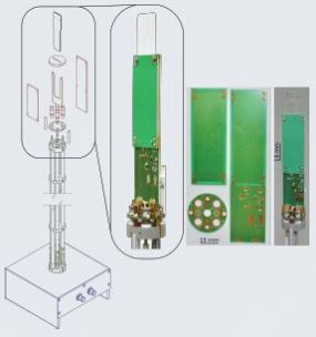
All microfluidic devices are produced from PMMA layers, bonded under heat and pressure. After initial sample loading, all experimental steps are carried out in situ inside the NMR spectrometer. Sample solutions (A & B, see Fig. 1) are contained within the microfluidic chip and the connecting reservoir capillary. Overall 40 ml of sample, equally divided into two solutions, is used. At the initial stage, only solution A is present inside the active sample circuit (Vs). A stepper-motor micropump (LabSmith) controls the injection volume (Vi) of sample B into the Vs. Mixing of the two volumes is achieved using a pneumatic microvalve [2] system to create a peristaltic flow [3] inside the Vs. NMR data are obtained from a 2.5 ml volume of the detection chamber by the double resonance Transmission Line Probe (TLP) [4], allowing for acquisition of proton-detected heteronuclear correlation spectra of proteins.
RESULTS AND DISCUSSION
Fig. 2 demonstrates the successful volume exchange of fumaric acid and DSS solutions inside the microfluidic device, as monitored by the intensity changes of the corresponding signals (Fig. 3). The concentrations were determined by calibration against a signal from sodium acetate, present in both solutions. The probe capacity for protein NMR is exemplified by the heteronuclear 15N and 13C spectra obtained for ubiquitin and the SH3 domain of human protein FYN (fynSH3) (Fig. 4). Experiments were carried out at 14.1T on 1 mM (ubiquitin) and 2 mM (fynSH3) samples with overall acquisition time of 400 min in each spectrum.
CONCLUSION
In this work we present a novel microfluidic platform for NMR detected protein-ligand studies. This system operates inside the NMR spectrometer, and allows for detailed control over sample injection and mixing, operating with a mixing volume of approx. 10 ml, and essentially zero dead volume.
ACKNOWLEDGMENTS
This project is jointly funded by the EU Horizon 2020 TISuMR project, and Institute for Life Sciences, University of Southampton, UK
Saraí M. Torres Delgado, Jan G. Korvink1 and Dario Mager*
Karlsruhe Institute of Technology – IMT, Eggenstein – Leopoldshafen, 76344, Germany
Tec.Nano 2019
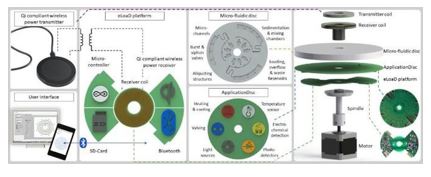
Over the last decade centrifugal microfluidic platforms have been of increasing interest for use in decentralized bioanalytical testing such as point-of-care diagnostics. This technology is particularly powerful due to the inherent ability to centrifuge samples like the ones required for blood processing. However, while the LoaD technique compared to LOC, has simplified basic operations such as valving, pumping, metering, mixing and sample preparation, solutions to other arising needs, such as the integration of (active) operations, or the readout of a bioassay result, has proven more challenging to achieve when the platform is under continuous rotation, a characteristic inherent to their working principle. As anyone can foresee, power and signal cables cannot be connected to a rotating system, because they will twist, entangled, and finally, disconnect or brake, hampering the integration of actuators and/or detectors into the system. These components are needed for a sensitive, reliable, time-independent, fast, direct and continuous interaction with the microfluidic disc while spinning and, thereby, enhancing the success of LoaD systems.
Hence, here we present the design and development of a low cost, compact and portable platform that co- rotates with the microfluidics disc, called the “electrified Lab-on-a-Disc (eLoaD) platform” which includes all modules necessary for it to be used in any diagnostic assay. Because it requires power, wireless energy transmission was introduced into the system. Hence, the platform was designed and fabricated to behave as a wireless power receiver compatible with the Qi standard, better known for its use in wireless charging of consumer electronic devices. Since most envisaged applications will require a control unit that provides enough computational power, a way to record data and real-time bidirectional communication between the user and the ongoing experiment, the platform comprises an Arduino microcontroller, an SD-Card and a Bluetooth module. The inclusion of those modules renders a flexible platform, easy to operate for most users with backgrounds ranging from biology to engineering and compatible with concurrently emerging trends and standard technologies.
As any laboratory that operates on basic and specialized equipment, the capabilities of the proposed system can be augmented by the addition of a second electronic board plug-compatible to the eLoaD. This additional board referred to as Application Disc in Fig. 1 contains the application-specific sensors and actuators. Such scheme leads to a higher degree of interaction and enables more sophisticated concepts to be implemented both in the control as well as in the readout. The performance of the platform was tested under several sensing and actuation experiments (1-3), some of which will be presented at the conference.
figure 1
Figure 1: Integration of the
wireless centrifugal system into conventional LoaD systems. A commercially
available Qi-compliant transmitter is inductively coupled to the eLoaD
platform. This fully integrated platform can control sensors and actuators
located on the Application Disc, which itself is simultaneously interacting
with the microfluidics disc. The disposable microfluidic disc and the reusable
Application Disc are typically designed for a particular application, whereas
the eLoaD platform, which implements the control logic, power, and
communication, is reused as a generic framework for all possible applications.
Interfacing of the eLoaD platform is enabled by Bluetooth communication, here
exemplary via an Android application program running on a portable device, and
from a PC running e.g. a LabVIEW script.
REFERENCES
(1) S. M. Torres Delgado et al., Lab Chip, vol. 16, no. 20, pp. 4002-4011, 2016.
(2) S. M. Torres Delgado et al., Biosensors and Bioelectronics, vol. 109, pp. 214 – 223, 2018.
Ruby E.H. Karsten, Nikolaas V.J.W. Krijnen, Maciej Grajewski, Elisabeth Verpoorte, Peter Olinga
Groningen Research Institute of Pharmacy, University of Groningen, Groningen, The Netherlands
Poster
SLAS Europe 2019
BelTox, Brussels 2019
Drug-induced
cholestasis (DIC), an adverse drug reaction, has a complex disease mechanism
with no good model for early
detection in drug development.
We are
developing an ex vivo mouse model
based on precision-cut liver slices (mPCLS) to study DIC. We incubate PCLS for 48h with glibenclamide
(a cholestatic drug). Cholestasis is ascertained by comparing control slices to
slices treated with drug, with and without an optimized bile acid (BA) mix. We studied
mPCLS viability and gene expression of BA transporters.
Non-toxic glibenclamide concentrations led to greater gene expression of basolateral and canalicular bile export transporters, the latter with a higher increase in the presence of BA mix. This study is the first that relates gene expression data to early DIC development in mPCLS. Once optimized, mPCLS will be incubated in a microfluidic device to monitor DIC onset in real time. We will use this model to elucidate disease mechanisms and perform drug toxicity screening.
1. de
Graaf IAM, Olinga P, de Jager MH, Merema MT, de Kanter R, van de Kerkhof EG,
e.a. Preparation and incubation of precision-cut liver and intestinal slices
for application in drug metabolism and toxicity studies. Nat Protoc. september
2010;5(9):1540–51.
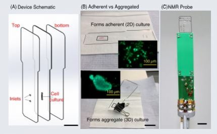
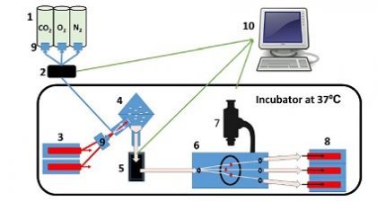
Time-lapse NMR spectroscopy was achieved up to 48 hours with MCF-7 cells, cultured in a microfluidic device of volume 2.5 l
Cells were cultured as an attached monolayer (2D) or an aggregated spheroid (3D) using same device design
Cell viability analysis was performed at the end of the NMR experiments
Manvendra Sharma, Bishnubrata Patra, William Hale and Marcel Utz
University of Southampton
ENC 2019
We present a novel microfluidic-NMR platform to combine high sensitivity NMR with microfluidics to perform generic NMR experiments on live systems under tightly controlled environment through microfluidics.
The current setup has already been used for a number of applications including microimaging of mouse liver tissue slices, heteronuclear 2D-NMR correlation experiments on proteins, parahydrogen induced hyperpolarisation, droplet NMR, reaction monitoring, and metabolic studies of mammalian cells.
Bishnubrata Patra, Manvendra Sharma, William Hale, Marcel Utz
University of Southampton
MicroTAS 2018
ABSTRACT
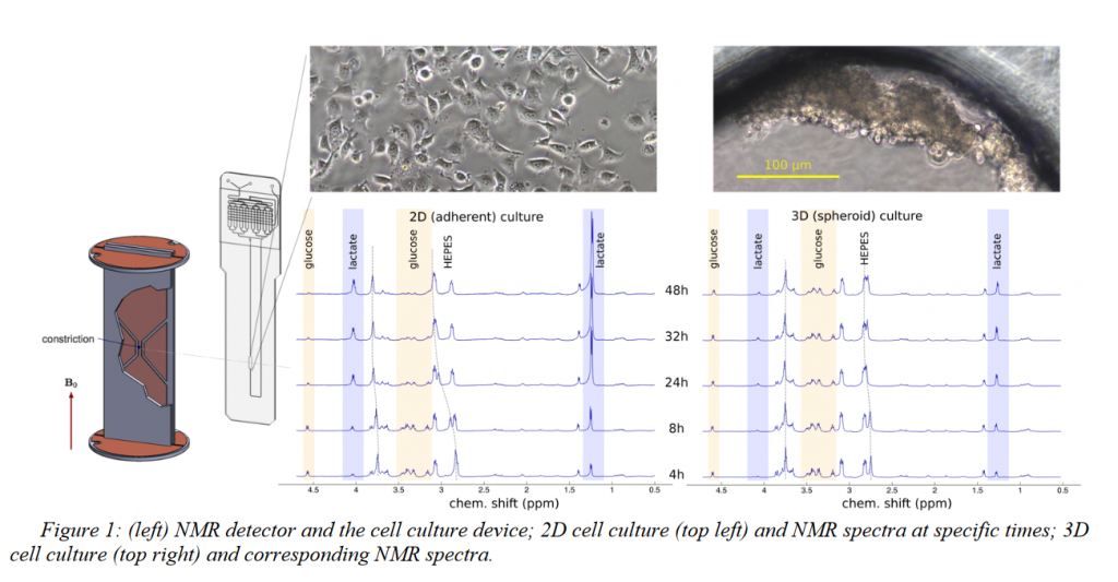
NMR metabolomics, the study of cellular metabolism, provides a new path to understand cellular response in vitro. In this article, we performed a real-time monitoring of adherent vs. spheroid cell culture using NMR metabolomics. The difference in lactate production provides a clear indication of different metabolic rates in spheroid compared to adherent cell culture. The developed microfluidic–NMR technique could be applicable towards real-time monitoring of a variety of ex-vivo biological samples, including tissue or organ on a chip.
Three-dimensional cell culture models (spheroids) provide valuable additional insight into the behavior of cancer cells since it mimics the features such as cell-cell contact and hindrance of metabolic diffusion which are not present in traditional cell culture.1On the other hand, NMR spectroscopy can provide in-situ monitoring of metabolomics even at microliter volume.2In this work, we used a novel integrated transmission-line NMR probe to directly compare the metabolomic activity of MCF-7 cells in adherent (2D) and spheroid (3D) culture. Microfluidic cell culture device of volume 2.5 μl volume was coated with fibronectin and with pluronic respectively to produce 2D and 3D cell cultures. Metabolomics observation was done up to 48 hours using NMR spectroscopy, followed by viability analysis. Clear differences in metabolomic activity were found, the spheroid (3D) showed much lower consumption of glucose, production of lactate, and acidosis compared to the attached (2D) culture.
EXPERIMENTAL
Microfluidic devices were made from thermally bonded PMMA layers. The cell culture chamber was coated overnight with fibronectin (1 mg/ ml) orpluronic (10 mg/ml). A suspension of 2.5 μl MCF-7 Cells (1000 cells/μl) in DMEM culture medium was introduced into each device. The devices were kept inside a cell culture incubator at 37° C and with 5% CO2. 1H NMR spectra were recorded using a 600 MHz spectrometerforboth2D and 3D cell culture devices and controls (without cells) periodically at 4, 8, 24, 32, and 48 hours. Cell viability was analyzed on the same devices using Live/Dead cell imaging kit [life technologies]and fluorescence microscopy.
RESULTS AND DISCUSSION
In this work, we used the equal number of cells with the equal volume of culture medium and the same culture environment to compare the metabolic activity of 2D vs.3D cultured MCF-7 cells by NMR spectroscopy. Our results showed that aggregated cells are metabolically less active and more viable after 48 hours compared to the attached cells
CONCLUSION
Figure 1 shows the 1HNMR spectra of 2D and 3D cultures up to 48 hours. 2D cells already started to produce more lactate compared to the 3D cell culture after 4 hours. At 48 hours 2D cell culture produces enough lactate to significantly shift the HEPES buffer resonances and consumed almost all the glucose. By contrast, 3D cells produced significantly less lactate. NMR spectra obtained in this way are of significant quality for unsupervised peak identification and metabolites present could be identified by mining the human metabolome database(figure 2). Figure 3 shows the viability analysis of 2D and Spheroid after 48 hours. The live stain (Green) indicates the healthy cells, whereas dead cells are stained as red. After 48 hours, 2D medium got enough acidified that cells are detaching, whereas cells survived strongly as spheroid.
ACKNOWLEDGEMENTS
This research project is funded by the horizon 2020 Framework program of the European Union.
REFERENCES
[1] F. Pampaloni, E. G. Reynaud, E.H.K. Stelzer, “The third dimension bridges the gap between cell culture and live tissue”. Nat Rev Mol Cell Biol. 8, 839, 2007
[2] G. Finch, A. Yilmaz, M. Utz, “An optimized detector for in-situ high-resolution NMR in microfluidic devices”. J. Mag. Res.262, 73-80, 2016
[3] D.S. Wishart, Y.D. Feunang, A. Marcu, A.C. Guo K. Liang, “ HMDB 4.0-The human metabolome database for 2018”. Nucleic Acids.Res.Jan 4; 4 6(D1): D608-17, 2018
Mazin Jouda, Erwin Fuhrer, Pedro Silva, Jan G. Korvink, and Neil MacKinnon
Karlsruhe Institute of Technology
ENC 2019
ABSTRACT
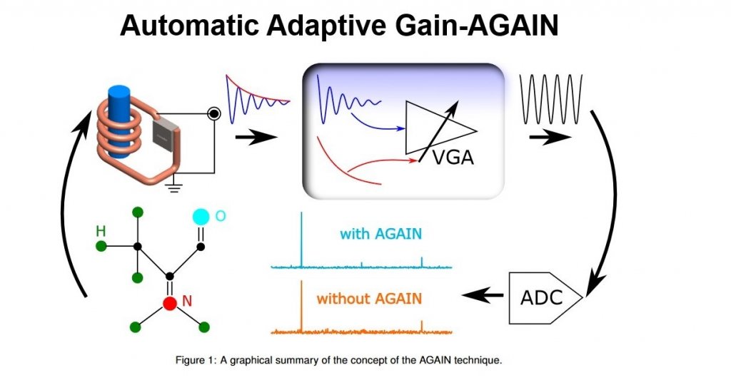
For an efficient NMR acquisition, an NMR spectrometer usually offers the option of automatically adjusting the receiver gain. The adjustment in this case is driven by the maximum amplitude of the FID, and once the receiver gain is set to a certain value, then it cannot be changed during the acquisition. But since the NMR signal is decaying by nature, only the first few points of the FID will be acquired with the full resolution of the analog-to-digital converter (ADC), whereas the rest of the FID will be acquired with a constantly decreasing resolution.
In this contribution we describe an innovative closed-loop control mechanism to automatically adapt the receiver gain over the acquisition time such that all of the FID points are digitized with the full ADC resolution [1]. A high performance digital lock-in amplifier was employed to extract the envelope of the FID, process it, and feed it back to control a variable-gain amplifier (VGA) inserted in the signal chain. As the FID decays the VGA gain and thus the overall receiver gain increases,even if the FID increases again due to beating for example, the VGA gain is adapted ensuring that no ADC overflow occurs.Additionally, the reported method includes a mechanism to precisely monitor the gain change over time. This is carried out via a pilot signal that propagates together with the NMR signal through the VGA. Knowledge of the gain modulation is of course essential to recover the NMR signal after the ADC stage.
The method, which we call AGAIN (Automatic Adaptive Gain), was verified experimentally. The results showed an SNR enhancement factor of 2.64 when AGAIN was applied to a custom spectrometer, while a maximum SNR enhancement factor of 1.45 was observed when applied to a commercial spectrometer. The AGAIN technique requires minimal additional hardware, which makes it general and thus can be readily combined with any commercial spectrometer.
REFERENCES
[1] M. Jouda, E. Fuhrer, P. Silva, J. G. Korvink, and N. MacKinnon. “Automatic adaptive gain for magnetic resonancesensitivity enhancement”. In:Analytical Chemistry (2019).